Systems Neuroscience & Neuroengineering
|
Research |

|
The RoLi lab is the joint lab of Drew Robson and Jennifer Li. The goal of the RoLi lab is to arrive at a set of design principles for cognition and neuromodulation in both biological and artificial systems.
We work with the larval zebrafish, a unique model organism with a compact and transparent brain in which nearly every neuron can be simultaneously recorded. Our neuroengineering team develops novel imaging systems to record and manipulate neural activity in freely swimming larval zebrafish, and our systems neuroscience team use these tools to gain a deeper understanding of the neural mechanisms that organize internal brain states and abstract cognitive representations. Our lab is a part of the Max Planck Institute for Biological Cybernetics |
|
Neuroengineering 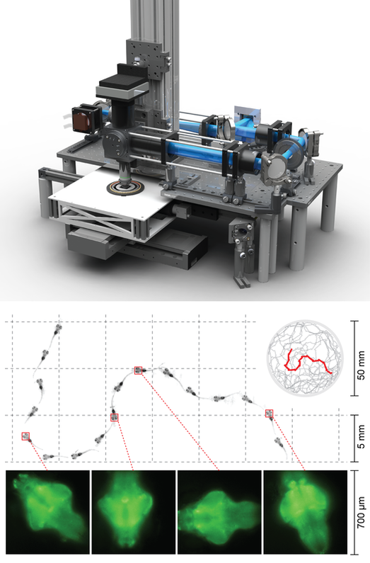
Kim DH, Kim J, Marques JC, Grama A, Hildebrand DGC, Gu W, Li JM* & Robson DN*. Pan-neuronal calcium imaging with cellular resolution in freely swimming zebrafish. Nature Methods. 2017 Nov;14(11):1107-1114
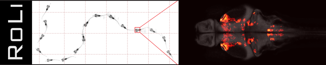
Over the last decade, larval zebrafish has emerged as an ideal system for brain-wide cellular resolution calcium imaging due to its small nervous system and high optical transparency. However, most existing imaging methods require the animal to be tethered under a microscope, which significantly restricts the range of behaviors the animal can perform. To overcome this limitation, our lab actively develops new microscopy systems that enable whole-brain imaging and manipulation in freely swimming larval zebrafish.
Using these systems, we are now investigating a wide repertoire of natural behaviors, including spatial navigation, social behavior, feeding, and reward. |
Systems Neuroscience 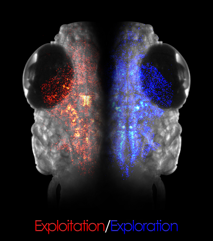
Marques JCǂ, Li Mǂ, Schaak D, Robson DN* & Li JM*. Internal state dynamics shape brainwide activity and foraging behaviour. Nature 2019. DOI: 10.1038/s41586-019-1858-z
The mammalian brain derived its basic architecture and organization from aquatic fish over 400 million years ago. However, it is far from clear the extent to which the wide array of mammalian cognitive processes and neuromodulatory states are already present in an ancestral form in non-mammalian vertebrate species, and how these ancestral networks may have constrained the evolutionary trajectory of the vertebrate brain.
Larval zebrafish is the smallest vertebrate animal model with demonstrated cognitive representations (e.g. abstract representation of space). Using advanced technologies pioneered by our engineering team, we investigate how the brain organizes internal states and cognitive representations, and create dynamical systems models for nonlinear neural dynamics in complex and recurrent neural networks (RNNs). Our current projects include:
|